Home
Uncategories
Blood Vessels Labeled Brain / Blood Vessels of the Brain & Spinal Cord - Atlas of Anatomy / In the article on the ventricles within the cns, we will discuss their structure and.
Blood Vessels Labeled Brain / Blood Vessels of the Brain & Spinal Cord - Atlas of Anatomy / In the article on the ventricles within the cns, we will discuss their structure and.
Blood Vessels Labeled Brain / Blood Vessels of the Brain & Spinal Cord - Atlas of Anatomy / In the article on the ventricles within the cns, we will discuss their structure and.. Blood travels from the heart in arteries, which branch into smaller and smaller vessels, eventually becoming arterioles. The blood vessels (and nerves) enter the brain through holes in the skull called foramina. If one of the major vessels becomes blocked, it is possible for blood flow to come across the circle of willis and prevent brain damage. Scientists are developing new strategies for attaching drugs to molecules naturally transported across the barrier (labeled in green and blue). Arterioles are small, nearly microscopic blood vessels that branch from muscular arteries.
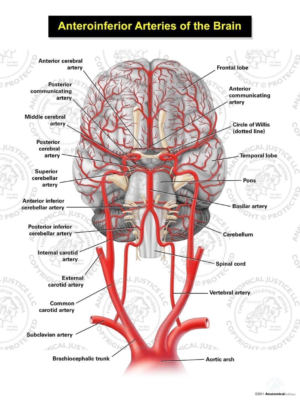
Blood is also supplied to the brain by the vertebral a. This vessel supplies blood to the front part of your brain, knows as your frontal lobe. The brain and its surrounding blood vessels exist in a close relationship. The carotid arteries and the vertebral arteries anterior cerebral artery (aca): He says the restricted vessels prevent the blood from draining fast enough and injure the brain by causing a build up of iron which leads to ms.
Anteroinferior Arteries of the Brain from anatomicaljustice.com Blood vessels examined in 3d allow scientists to examine circulation in the brain giving greater understanding of how dementia, brain cancer and stroke may affect veins and capillaries in this important organ. Blood supply to the brain is supplied by two main pairs of arteries, the internal carotid arteries and the vertebral arteries. Arterioles are small, nearly microscopic blood vessels that branch from muscular arteries. The smallest arterioles consist of endothelium surrounded by a single layer of smooth muscle. Only some of the vessels that exist in a real brain have been labeled. Function and homeostasis of the brain relies on communication between its complex network of cells. • identification of blood vessels as arteries, capillaries or veins from the structure of their walls. There is a right sided aca and a left sided aca.
Scientists are developing new strategies for attaching drugs to molecules naturally transported across the barrier (labeled in green and blue).
| study material, lecturing notes, assignment mr angiography makes the use of the mri magnetfor the examination of the blood vessels in this test no catheter is required to be introduced in the blood. Label the blood vessels of the male pelvis using the hints provided. Supplies the posterior brain, blood supply to the entire brain is ensured by anastomoses between the vessels. Blood flows throughout the body tissues in blood vessels, via bulk flow (i.e., all constituents together and in one direction). Label the veins of the anterior forearm and hand. Researchers have discovered how cells of the blood vessels sense the metabolic condition of the brain and alter vascular function in response. Vessel clusters lying within each region were labeled accordingly. They also take waste and carbon dioxide away from the tissues. Blood in the brain is supplied by two pairs of large blood vessels (arteries): The carotid arteries and the vertebral arteries anterior cerebral artery (aca): Blood vessels examined in 3d allow scientists to examine circulation in the brain giving greater understanding of how dementia, brain cancer and stroke may affect veins and capillaries in this important organ. Instead, transport is controlled mostly by the dilation of vessels. Blood vessels in red in close communication with proliferating neuronal cells in the mouse cortex at embryonic day 10.
Supplies the posterior brain, blood supply to the entire brain is ensured by anastomoses between the vessels. Label the blood vessels of the male pelvis using the hints provided. If there are blockages in blood vessels or if blood doesn't reach the brain for some reason, for how long can the brain maintain itself without oxygen? Label the blood vessels in the inferior view of the brain using the hints provided. Identify all of the blood vessels that are illustrated in the figure as you can while holding or otherwise examining whole brain specimens.
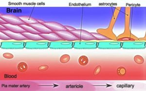
Bloodflow and Function of the Left Middle Cerebral Artery Territory from anatomicaljustice.com This is particularly important structure due to its clinical implications, which are discussed in more detail in the article. Blood travels from the heart in arteries, which branch into smaller and smaller vessels, eventually becoming arterioles. The difference in the structural characteristics of arteries, capillaries and veins is attributable to their respective functions. Vessel clusters lying within each region were labeled accordingly. In healthy adults, the walls of blood vessels are so tight that foreign bodies cannot reach the brain. The vessels allow blood to be pumped at a high pressure to deliver nutrients and. The dense tight junctions between endothelial cells prevent paracellular transport through the. Fill in the blanks with the appropriate words to describe blood flow from the heart.
Identify all of the blood vessels that are illustrated in the figure as you can while holding or otherwise examining whole brain specimens.
⇒ click on the diagram to show / hide labels. Blood travels from the heart in arteries, which branch into smaller and smaller vessels, eventually becoming arterioles. Examine a second specimen and notice any differences, such as asymmetries in the size of the vertebral or posterior communicating arteries. The dense tight junctions between endothelial cells prevent paracellular transport through the. If one of the major vessels becomes blocked, it is possible for blood flow to come across the circle of willis and prevent brain damage. Scientists are developing new strategies for attaching drugs to molecules naturally transported across the barrier (labeled in green and blue). The brain and its surrounding blood vessels exist in a close relationship. Blood vessels are tubes that run through the transport system in which blood is transported. As well as providing new insights into the. Supplies the anterior brain and the vertebral a. Diseases of the brain and nervous system(a health education guide): These vessels of the brain supply blood to the brain stem, the rear of the cerebrum and part of the cerebellum. In healthy adults, the walls of blood vessels are so tight that foreign bodies cannot reach the brain.
These vessels of the brain supply blood to the brain stem, the rear of the cerebrum and part of the cerebellum. Traditionally, pais has been explained as being caused by a blood clot forming within the ageing placenta. Blood supply to the brain is supplied by two main pairs of arteries, the internal carotid arteries and the vertebral arteries. Instead, transport is controlled mostly by the dilation of vessels. This is particularly important structure due to its clinical implications, which are discussed in more detail in the article.
Different Triggers - Different Illnesses: Exercise Affects Brains of ME/CFS and GWI Patients ... from www.healthrising.org Neurosciencenews.com image is adapted from the university of surrey press release. To explore spatial variations in the venous pulsatility characteristics, vessels were considered according to (2018). Blood flows throughout the body tissues in blood vessels, via bulk flow (i.e., all constituents together and in one direction). Comes off the subclavian a., ascends although the internal carotid a. Blood vessels examined in 3d allow scientists to examine circulation in the brain giving greater understanding of how dementia, brain cancer and stroke may affect veins and capillaries in this important organ. The difference in the structural characteristics of arteries, capillaries and veins is attributable to their respective functions. The dense tight junctions between endothelial cells prevent paracellular transport through the. The carotid arteries and the vertebral arteries anterior cerebral artery (aca):
Scientists are developing new strategies for attaching drugs to molecules naturally transported across the barrier (labeled in green and blue).
Blood vessels and lymph nodes. The blood vessels are the components of the circulatory system that transport blood throughout the human body. The dense tight junctions between endothelial cells prevent paracellular transport through the. Arterioles are small, nearly microscopic blood vessels that branch from muscular arteries. Internal carotid artery (anterior circulation), vertebral artery (posterior circulation), and their hexagonal anastomotic network called blood brain barrier refers to the wall between the brain tissue and blood vessels. The difference in the structural characteristics of arteries, capillaries and veins is attributable to their respective functions. The smallest arterioles consist of endothelium surrounded by a single layer of smooth muscle. This is particularly important structure due to its clinical implications, which are discussed in more detail in the article. But as we age, the walls loosen, which may allow amyloid beta and tau proteins to. Label the blood vessels of the male pelvis using the hints provided. Label the blood vessels in the inferior view of the brain using the hints provided. Fill in the blanks with the appropriate words to describe blood flow from the heart. These vessels transport blood cells, nutrients, and oxygen to the tissues of the body.
Instead, transport is controlled mostly by the dilation of vessels blood vessels labeled. Vessel clusters lying within each region were labeled accordingly.

0 Comments:
Posting Komentar